
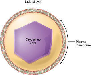
Peroxisomes are found in both plant and animal cells, they were the of the last organelle to be discovered and characterised in eukaryotic cells (Hu et al. 2012). They have a simple structure consisting of two major parts; an outer later, single lipid-bilayer membrane and a crytalline core. The image to the left (taken from boundless.com) shows the basic structure of the peroxisome with a lipid bilayer surrounded by a plasma membrane and a condensed crystalline core in the center. The core contains a variety of enzymes for catalysis of cell functions, mainly metabolism. Typically, the most concentrated of these enzymes, catalase or urate oxidase, can become so high in number that they aggregate to form a crystalline core in the centre of the organelle. This structure can be seen in electron micrographs.
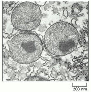
The image to the right shows an electron micrograph of peroxisomes. The three circular structures are peroxisomes and in the two lower ones a dark central region can be seen. In this example the peroxisomes contain a highly condensed urate oxidase crystalline core.The enzymes are accumulated in large numbers so once they are needed in the cell they can be released quickly so the peroxisomes can act rapidly when required. Once a peroxisome has formed as a result of biogenesis it needs to increase in size. this is done by the post-translational import of newly synthesized proteins from the cytosol which are transported across the peroxisome membrane and into the core. several of the proteins are kept in the membrane (Lazarow 1995).
Image taken from (Schrader and Fahimi 2008)
